The gnathostomes, or jawed vertebrates (Infraphylum Gnathostomata, Subphylum Vertebrata) comprise all living vertebrates except for the Cyclostomata.
The stem group of the Gnathostomes is very well represented in the fossil record, and its general phylogeny is generally accepted to have the form shown below:
The stem group of the Gnathostomes is very well represented in the fossil record, and its general phylogeny is generally accepted to have the form shown below:

Figure 1. Summary phylogenetic tree of the stem-Gnathostomata
Each of the terminal taxa shown in the above tree, with the exception of the placoderms, are clades with a common ancestor. They represent dead-end side-branches of the gnathostome stem line and contain only jawless species. The placoderms, on the other hand, are paraphyletic because they do not contain all the descendants of their last common ancestor (LCA); they share that LCA with the gnathostome crown group.
In this section we will consider each of the side branches of Figure 1 separately, using pruned versions of phylogenetic trees taken from the literature. Pruning is necessary because many of the fossils included in the published trees lie far out on the side branches shown in Figure 1, phylogenetically distant from the main stem line leading to the crown group; these are less informative as transitional fossils than those that lie close to the main stem line, and are thus excluded from consideration here.
Pteraspidomorphi
The Pteraspidomorphi, also known as the Pteraspidomorpha, represent the most basal members of the stem gnathostomes. They contain the oldest known stem-group fossils:
One version of the phylogeny of the pteraspidomorphs (constructed using the R package "strap" (Bell and Lloyd, 2015) and selecting the "equal" option that requires specification of the age of the root of the tree), pruned to exclude the species furthest from the basal node, is shown below:
In this section we will consider each of the side branches of Figure 1 separately, using pruned versions of phylogenetic trees taken from the literature. Pruning is necessary because many of the fossils included in the published trees lie far out on the side branches shown in Figure 1, phylogenetically distant from the main stem line leading to the crown group; these are less informative as transitional fossils than those that lie close to the main stem line, and are thus excluded from consideration here.
Pteraspidomorphi
The Pteraspidomorphi, also known as the Pteraspidomorpha, represent the most basal members of the stem gnathostomes. They contain the oldest known stem-group fossils:
- Arandaspis prionotolepis, a primitive jawless fish, belonging to the Arandaspida (the sister group of the Heterostraci), from the Middle Ordovician Stairway Sandstone of Mount Watt and Mount Charlotte, Northern Territory, Australia (Ritchie and Gilbert-Tomlinson, 1977; Benton et al, 2015).
- Sacabambaspis janvieri, another jawless fish belonging to the Arandaspida and found in the Middle Ordovician Anzaldo Formation at Sacabamba in the department of Cochabamba, Bolivia (Gagnier et al, 1986; Sallan et al, 2018).
One version of the phylogeny of the pteraspidomorphs (constructed using the R package "strap" (Bell and Lloyd, 2015) and selecting the "equal" option that requires specification of the age of the root of the tree), pruned to exclude the species furthest from the basal node, is shown below:

Figure 2. Time tree of the Pteraspidomorphi. Root age specified as 508.5 million years.
Species from the above tree for which images are available in the public domain are shown below (click on image for a larger view; this applies to all fossil image sets further below on this page):


Names in red indicate that the fossil is younger than the oldest known crown-group fossil.
Figure 3. Images of stem-Gnathostomata (Pteraspidomorphi)
The above images, together with those in the following image sets for the other taxa of the stem gnathostomes, are placed in left-to-right order from the most basal to the furthest crownward, according to the topology of the phylogenetic tree. No obvious trends can be seen; all the fossils have a generally similar appearance.
The stem line belonging to the Pteraspidomorphi includes a ghost lineage (shown as a blue bar in Figure 1), because the oldest known member of the stem-Cyclostomata (Cambropustula kinnekullensis, of Late Cambrian age; see page on the Cyclostomata) is at least 30 million years older than the stem-Gnathostomata, which are of Middle Ordovician and younger age. Given that the two stem groups must have appeared at the same time, the gnathostome stem-group transition must also have begun in the Late Cambrian.
Thelodonti
A possible phylogeny of the Thelodonti is shown in the time tree below:

Figure 4. Time tree of the Thelodonti
Species from the above tree with public-domain images are shown below:


Names in red indicate that the fossil is younger than the oldest known crown-group fossil.
Figure 5. Images of stem-Gnathostomata (Thelodonti)
These species are, like the pteraspidomorphs, jawless and armored, but they do not show any clear trend through the series shown above. The armor of the thelodonts comprises scales, but these were generally dispersed after death, such that their anatomy is poorly understood (Donoghue and Smith, 2001). However, one apparent difference between the thelodonts and the pteraspidomorphs is that the latter have a more sharply-differentiated head.
One species, Pezopallichthys ritchiei, shows little resemblance to the other fossils shown above. It appears to have a vertically-elongated body rather than the flat form exhibited by the other species. However, the scales of Pezopallichthys have a morphology that demonstrates that the species belongs to the thelodonts (Wilson & Märss, 2009; Ferrón & Botella, 2017).
One species, Pezopallichthys ritchiei, shows little resemblance to the other fossils shown above. It appears to have a vertically-elongated body rather than the flat form exhibited by the other species. However, the scales of Pezopallichthys have a morphology that demonstrates that the species belongs to the thelodonts (Wilson & Märss, 2009; Ferrón & Botella, 2017).
Galeaspida
The time tree below represents a possible phylogeny of the Galeaspida:
The time tree below represents a possible phylogeny of the Galeaspida:

Figure 6. Time tree of the Galeaspida
This clade, illustrated by the following images, comprises species that all have massive head-shields:


Names in red indicate that the fossil is younger than the oldest known crown-group fossil.
Figure 7. Images of stem-Gnathostomata (Galeaspida)
The shape and nature of the head-shield is superficially similar to the shield of the Heterostraci clade of the pteraspidomorphs (for example, Hibernaspis and Olbiaspis in Figure 3).
Osteostraci
The next clade to separate from the gnathostome stem line was the Osteostraci. A version of their phylogeny is illustrated below:
Osteostraci
The next clade to separate from the gnathostome stem line was the Osteostraci. A version of their phylogeny is illustrated below:

Figure 8. Time tree of the Osteostraci
The osteostracans are generally similar to the galeaspids, but their bone type is cellular (Janvier, 1984) while at least part of the bone in the galeaspids is acellular (Keating et al, 2018) and the bone structure of the head differs in detail. Here are some examples:


Names in red indicate that the fossil is younger than the oldest known crown-group fossil.
Figure 9. Images of stem-Gnathostomata (Osteostraci)
Most of the species shown above (e.g. Cephalaspis) have a head-shield with a characteristic pointed lateral extension on each side.
Placoderms
The time tree shown below represents a possible topology of the phylogenetic relationships between the placoderms, which are the jawed stem gnathostomes (Zhu et al, 2016):
Placoderms
The time tree shown below represents a possible topology of the phylogenetic relationships between the placoderms, which are the jawed stem gnathostomes (Zhu et al, 2016):

Figure 10. Time tree of the placoderms
The species shown above for which public-domain images are available are illustrated below:


Names in red indicate that the fossil is younger than the oldest known crown-group fossil.
Figure 11. Images of stem-Gnathostomata (Placoderms)
One of the most basal Placoderm clades, the Antiarchi, differ from more basal stem gnathostomes in being more heavily armored, with a box-like thoracic shield and elongate pectoral fins encased in bone (Zhu et al, 2012). A good example of the bony pectoral fins can be seen above in the antiarchs Yunnanolepis, Bothriolepis, Microbrachius, Pterichthyodes and Remigolepis.
A more crownward Placoderm clade, the Arthrodira, differs from more basal stem-Gnathostomata in having a movable joint between the head and the body armor. Like the antiarchs, they also had teeth (Rücklin et al, 2012). The arthrodires are represented in the above tree (Fig. 10) by Dicksonosteus, Groenlandaspis, Holonema, Kujdanowiaspis, Wuttagoonaspis and Cowralepis.
Summary of the stem-Gnathostomata
In summary, all the stem gnathostomes were armored fishes, although the nature of the armor - whether consisting of plates or scales - varied through the development of the stem group. Furthermore, the fossil record shows a transition from jawless fishes without teeth to jawed fish with teeth. Other events in the development of the stem group were the appearance of paired fins and articulation between the head and the body.
Transition to the crown group
Some idea of the nature of the transition from the stem group to the crown group of the gnathostomes can be derived from a comparison of the above images with the examples of early crown-Gnathostomata shown below:
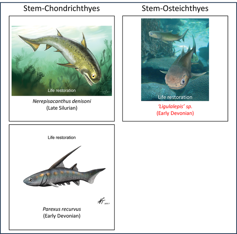
Names in red indicate that the fossil is younger than the oldest known crown-group fossil.
Figure 12. Examples of early crown-Gnathostomata
It is noteworthy that the crown gnathostomes shown above, which appear quite similar to modern fishes, are significantly different in general appearance, according to the life restorations shown above, from many of the placoderms, which are the most-crownward stem gnathostomes.
Page last updated July 6th, 2026.
References
Bell, M. A., & Lloyd, G. T. (2015). strap: an R package for plotting phylogenies against stratigraphy and assessing their stratigraphic congruence. Palaeontology, Vol. 58, No. 2, pp. 379-389.
Benton, M. J., Donoghue, P. C., Asher, R. J., Friedman, M., Near, T. J., & Vinther, J. (2015). Constraints on the timescale of animal evolutionary history. Palaeontologia Electronica, 18(1), 1-106.
Brazeau, M. D., Castiello, M., El Fassi El Fehri, A., Hamilton, L., Ivanov, A. O., Johanson, Z., & Friedman, M. (2023). Fossil evidence for a pharyngeal origin of the vertebrate pectoral girdle. Nature, 623(7987), 550-554.
Donoghue, P. C., & Smith, M. P. (2001). The anatomy of Turinia pagei (Powrie), and the phylogenetic status of the Thelodonti. Earth and Environmental Science Transactions of The Royal Society of Edinburgh, 92(1), 15-37.
Ferrón, H. G., & Botella, H. (2017). Squamation and ecology of thelodonts. PloS one, 12(2), e0172781.
Ferrón, H. G., Martínez‐Pérez, C., Turner, S., Manzanares, E., & Botella, H. (2018). Patterns of ecological diversification in thelodonts. Palaeontology, 61(2), 303-315.
Gagnier, P. Y., Blieck, A. R., & Rodrigo, G. (1986). First Ordovician vertebrate from South America. Geobios, 19(5), 629-634.
Janvier, P. (1984). The relationships of the Osteostraci and Galeaspida. Journal of Vertebrate Paleontology, 4(3), 344-358.
Keating, J. N., Marquart, C. L., Marone, F., & Donoghue, P. C. (2018). The nature of aspidin and the evolutionary origin of bone. Nature ecology & evolution, 2(9), 1501-1506.
Randle, E., Keating, J. N., & Sansom, R. S. (2025). A phylogeny for Heterostraci (stem‐gnathostomes). Papers in Palaeontology, 11(5), e70030.
Ritchie, A., & Gilbert-Tomlinson, J. (1977). First Ordovician vertebrates from the southern hemisphere. Alcheringa, 1(4), 351-368.
Rücklin, M., Donoghue, P. C., Johanson, Z., Trinajstic, K., Marone, F., & Stampanoni, M. (2012). Development of teeth and jaws in the earliest jawed vertebrates. Nature, 491(7426), 748-751.
Sallan, L., Friedman, M., Sansom, R. S., Bird, C. M., & Sansom, I. J. (2018). The nearshore cradle of early vertebrate diversification. Science, 362(6413), 460-464.
Sansom, R. S., Randle, E., & Donoghue, P. C. (2015). Discriminating signal from noise in the fossil record of early vertebrates reveals cryptic evolutionary history. Proceedings of the Royal Society B: Biological Sciences, 282(1800).
Wilson, M. V., & Märss, T. (2009). Thelodont phylogeny revisited, with inclusion of key scale-based taxa. Estonian Journal of Earth Sciences, 58(4).
Zhang, Y., Shan, X., Lin, X., Gai, Z., & Donoghue, P. C. (2026). A New Dayongaspid Galeaspid from the Silurian of the Lower Yangtze Region: Implications for Biogeography and the Evolution of Key Adaptations in Galeaspids. Journal of Earth Science, 37(1), 303-316.
Zhu, M., Yu, X., Choo, B., Wang, J., & Jia, L. (2012). An antiarch placoderm shows that pelvic girdles arose at the root of jawed vertebrates. Biology Letters, 8(3), 453-456.
Zhu, M., Ahlberg, P. E., Pan, Z., Zhu, Y., Qiao, T., Zhao, W., ... & Lu, J. (2016). A Silurian maxillate placoderm illuminates jaw evolution. Science, 354(6310), 334-336.
Zhu, Y. A., Li, Q., Lu, J., Chen, Y., Wang, J., Gai, Z., ... & Zhu, M. (2022). The oldest complete jawed vertebrates from the early Silurian of China. Nature, 609(7929), 954-958.
Image credits - Stem-Gnathostomes
- Figure 3 (Eriptychius americanus): Richard P. Dearden, Agnese Lanzetti, Sam Giles, Zerina Johanson, Andy S. Jones, Stephan Lautenschlager, Emma Randle & Ivan J. Sansom, CC BY 4.0 <https://creativecommons.org/licenses/by/4.0>, via Wikimedia Commons
- Figure 3 (headshield of Astraspis desiderata): L. Beverly Tarlo, CC BY-SA 4.0 <https://creativecommons.org/licenses/by-sa/4.0>, via Wikimedia Commons
- Figure 3 (Astraspis desiderata, life restoration): Nobu Tamura, under Creative Commons Attribution- ShareAlike (CC BY-SA) licence
- Figure 3 (Arandaspis prionotolepis): Nobu Tamura, under a Creative Commons 3.0 Unported (CC BY-NC-ND 3.0) license
- Figure 3 (Sacabambaspis janvieri, fossils): Ghedoghedo / CC BY-SA (https://creativecommons.org/licenses/by-sa/3.0)
- Figure 3 (Sacabambaspis janvieri, life restoration): Nobu Tamura, under Creative Commons Attribution- ShareAlike (CC BY-SA) licence
- Figure 3 (Olbiaspis coalescens): Apokryltaros, CC BY-SA 4.0 <https://creativecommons.org/licenses/by-sa/4.0>, via Wikimedia Commons
- Figure 3 (Hibernaspis macrolepis): Nobu Tamura, licensed under Creative Commons Attribution- ShareAlike (CC BY-SA)
- Figure 5 (Archipelepis turbinata): Nobu Tamura, licensed under Creative Commons Attribution- ShareAlike (CC BY-SA) license
- Figure 5 (Turinia pagei): Nobu Tamura, licensed under Creative Commons Attribution- ShareAlike (CC BY-SA) license
- Figure 5 (Thelodus parvidens): Nobu Tamura, licensed under Creative Commons Attribution- ShareAlike (CC BY-SA) license
- Figure 5 (Lanarkia spinosa): Ghedoghedo, CC BY-SA 4.0 <https://creativecommons.org/licenses/by-sa/4.0>, via Wikimedia Commons
- Figure 5 (Lanarkia horrida): Nobu Tamura, licensed under Creative Commons Attribution- ShareAlike (CC BY-SA) license
- Figure 5 (Pezopallichthys ritchei): Nobu Tamura, licensed under Creative Commons Attribution- ShareAlike (CC BY-SA) license
- Figure 7 (Hanyangaspis guodingshanensis): Nobu Tamura, licensed under Creative Commons Attribution- ShareAlike (CC BY-SA) license
- Figure 7 (Dayongaspis hunanensis): Apokryltaros, CC BY-SA 4.0 <https://creativecommons.org/licenses/by-sa/4.0>, via Wikimedia Commons
- Figure 7 (Tujiaaspis sp.): Kylinxia, CC BY-SA 4.0 <https://creativecommons.org/licenses/by-sa/4.0>, via Wikimedia Commons
- Figure 7 (Shuyu zhejiangensis): Nobu Tamura, licensed under Creative Commons Attribution- ShareAlike (CC BY-SA) license
- Figure 9 (Hemicyclaspis sp.): Haplochromis, CC BY-SA 3.0 <https://creativecommons.org/licenses/by-sa/3.0>, via Wikimedia Commons
- Figure 9 (Hemicyclaspis murchisoni): Nobu Tamura, licensed under Creative Commons Attribution- ShareAlike (CC BY-SA) license
- Figure 9 (Ateleaspis tesselata): Nobu Tamura, licensed under Creative Commons Attribution- ShareAlike (CC BY-SA) license
- Figure 9 (Cephalaspis sp.): Haplochromis, CC BY-SA 3.0 <https://creativecommons.org/licenses/by-sa/3.0>, via Wikimedia Commons
- Figure 9 (Cephalaspis lyelli): Nobu Tamura, licensed under Creative Commons Attribution- ShareAlike (CC BY-SA) license
- Figure 9 (Wladysagitta janvieri): Nobu Tamura, licensed under Creative Commons Attribution- ShareAlike (CC BY-SA) license
- Figure 9 (Parameteoraspis oberon): MNHN, CC BY 4.0 <https://creativecommons.org/licenses/by/4.0>, via Wikimedia Commons
- Figure 11 (Brindabellaspis stensioi): Entelognathus, CC BY-SA 4.0 <https://creativecommons.org/licenses/by-sa/4.0>, via Wikimedia Commons
- Figure 11 (Kolymaspis sibirica): Open access article by Brazeau, M. D., Castiello, M., El Fassi El Fehri, A., Hamilton, L., Ivanov, A. O., Johanson, Z., & Friedman, M. (2023). Fossil evidence for a pharyngeal origin of the vertebrate pectoral girdle. Nature, 623(7987), 550-554.
- Figure 11 (Skull of Romundina stellina): Open Access article by Dupret, V., Sanchez, S., Goujet, D., & Ahlberg, P. E. (2017). The internal cranial anatomy of Romundina stellina Ørvig, 1975 (Vertebrata, Placodermi, Acanthothoraci) and the origin of jawed vertebrates—Anatomical atlas of a primitive gnathostome. PLoS One, 12(2), e0171241.
- Figure 11 (Romundina stellina, life restoration): Apokryltaros / CC BY-SA (https://creativecommons.org/licenses/by-sa/3.0)
- Figure 11 (Yunnanolepis chii): Nobu Tamura, under Creative Commons Attribution- ShareAlike (CC BY-SA) license
- Figure 11 (Parayunnanolepis xitunensis, fossil): Open Access article by Wang, Y., & Zhu, M. (2022). Squamation and scale morphology at the root of jawed vertebrates. Elife, 11, e76661.
- Figure 11 (Parayunnanolepis xitunensis, life restoration): Open Access article by Wang, Y., & Zhu, M. (2022). Squamation and scale morphology at the root of jawed vertebrates. Elife, 11, e76661.
- Figure 11 (Gemuendina stuertzi, fossil): Bloopityboop, CC BY-SA 4.0 <https://creativecommons.org/licenses/by-sa/4.0>, via Wikimedia Commons
- Figure 11 (Gemuendina stuertzi, life restoration): Nobu Tamura, under Creative Commons Attribution- ShareAlike (CC BY-SA) license
- Figure 11 (Pterichthyodes milleri, fossil): Daderot, CC0, via Wikimedia Commons
- Figure 11 (Pterichthyodes milleri, life restoration): Nobu Tamura, under Creative Commons Attribution- ShareAlike (CC BY-SA) license
- Figure 11 (Head and pectoral appendages of Microbrachius dicki): Orkney Landscapes, under Attribution-NonCommercial-ShareAlike 2.0 licence
- Figure 11 (Microbrachius dicki, life restoration): Fossiilid.info under CC BY 4.0 Licence
- Figure 11 (Bothriolepis canadensis, fossil): Hectonichus, CC BY-SA 3.0 <https://creativecommons.org/licenses/by-sa/3.0>, via Wikimedia Commons
- Figure 11 (Bothriolepis canadensis, life restoration): Nobu Tamura, under Creative Commons Attribution- ShareAlike (CC BY-SA) license
- Figure 11 (Xiushanosteus mirabilis): Kylinxia, CC BY-SA 4.0 <https://creativecommons.org/licenses/by-sa/4.0>, via Wikimedia Commons
- Figure 11 (Jagorina pandora): Apokryltaros at English Wikipedia, CC BY-SA 3.0 <http://creativecommons.org/licenses/by-sa/3.0/>, via Wikimedia Commons
- Figure 11 (Remigolepis walkeri): Sébastien Olive, CC BY 4.0 <https://creativecommons.org/licenses/by/4.0>, via Wikimedia Commons
- Figure 11 (Entelognathus primordialis, fossil): Xindong Cui, Matt Friedman, Yilun Yu, You-an Zhu & Min Zhu, CC BY 4.0 <https://creativecommons.org/licenses/by/4.0>, via Wikimedia Commons
- Figure 11 (Entelognathus primordialis, life restoration): Xindong Cui, Matt Friedman, Yilun Yu, You-an Zhu & Min Zhu, CC BY 4.0 <https://creativecommons.org/licenses/by/4.0>, via Wikimedia Commons
- Figure 11 (Janusiscus schultzei): Fossiilid.info under CC BY 4.0 Licence
- Figure 11 (Diandongpetalichthys liaojiaoshanensis): Apokryltaros, CC BY-SA 3.0 <https://creativecommons.org/licenses/by-sa/3.0>, via Wikimedia Commons
- Figure 11 (Dicksonosteus arcticus, fossil): Ghedo, CC BY-SA 4.0 <https://creativecommons.org/licenses/by-sa/4.0>, via Wikimedia Commons
- Figure 11 (Dicksonosteus arcticus, life restoration): Nobu Tamura, under Creative Commons Attribution- ShareAlike (CC BY-SA) license
- Figure 11 (Preserved spinal plate and thoradic shield of Kujdanowiaspis sp.): Entelognathus, CC BY-SA 4.0 <https://creativecommons.org/licenses/by-sa/4.0>, via Wikimedia Commons
- Figure 11 (Kujdanowiaspis sp., life restoration): Entelognathus, CC BY-SA 4.0 <https://creativecommons.org/licenses/by-sa/4.0>, via Wikimedia Commons
- Figure 11 (Quasipetalichthys haikouensis): Apokryltaros at English Wikipedia, CC BY-SA 3.0 <http://creativecommons.org/licenses/by-sa/3.0/>, via Wikimedia Commons
- Figure 11 (Groenlandaspis howittensis, fossils): Open Access article by Fitzpatrick, A. N., Clement, A. M., & Long, J. A. (2024). Unique dental arrangement in a new species, Groenlandaspis howittensis (Placodermi, Arthrodira) from the Middle Devonian of Mount Howitt, Victoria, Australia. PeerJ, 12, e18759.
- Figure 11 (Groenlandaspis howittensis, life restoration): Open Access article by Fitzpatrick, A. N., Clement, A. M., & Long, J. A. (2024). Unique dental arrangement in a new species, Groenlandaspis howittensis (Placodermi, Arthrodira) from the Middle Devonian of Mount Howitt, Victoria, Australia. PeerJ, 12, e18759.
- Figure 11 (Holonema westolli): Apokryltaros at English Wikipedia, CC BY-SA 3.0 <https://creativecommons.org/licenses/by-sa/3.0>, via Wikimedia Commons
- Figure 11 (Headshield of Wuttagoonaspis fletcheri): Fossiilid.info under CC BY 4.0 Licence
- Figure 11 (Wuttagoonaspis fletcheri, life restoration): Apokryltaros, CC BY-SA 3.0 <https://creativecommons.org/licenses/by-sa/3.0>, via Wikimedia Commons
- Figure 11 (Lunaspis broili): Ghedoghedo, Public domain, via Wikimedia Commons
- Figure 11 (Partial head of Macropetalichthys rapheidolabis): James St. John, CC BY 2.0 <https://creativecommons.org/licenses/by/2.0>, via Wikimedia Commons
- Figure 11 (Macropetalichthys rapheidolabis, life restoration): ДиБгд, CC BY 4.0 <https://creativecommons.org/licenses/by/4.0>, via Wikimedia Commons
- Figure 11 (Cowralepis mclachlani, fossil): Oilshale, CC BY-SA 4.0 <https://creativecommons.org/licenses/by-sa/4.0>, via Wikimedia Commons
- Figure 11 (Cowralepis mclachlani, life restoration): Apokryltaros, CC BY-SA 3.0 <https://creativecommons.org/licenses/by-sa/3.0>, via Wikimedia Commons
- Figure 12 (Nerepisacanthus denisoni): Danielle Dufault [CC BY 2.5 (https://creativecommons.org/licenses/by/2.5)]
- Figure 12 (Parexus recurvus): Nobu Tamura, licensed under Creative Commons Attribution- ShareAlike (CC BY-SA)
- Figure 12 ('Ligulalepis', life restoration): Open Access article Clement, A. M., King, B., Giles, S., Choo, B., Ahlberg, P. E., Young, G. C., & Long, J. A. (2018). Neurocranial anatomy of an enigmatic Early Devonian fish sheds light on early osteichthyan evolution. Elife, 7, e34349