The amniote clade (within Superclass Tetrapoda) represents animals that produce an air-breathing egg with protective membranes that allow the egg to be laid on land or gestated within the mother. The amniotes comprise two clades: the mammals and the saurians (reptiles and birds). Mammals are synapsids, having only one hole in the skull behind each eye, while saurians are diapsids, with two holes (except for the turtles, or Testudines, in which both holes behind the eye are thought to have closed up during the evolutionary process; Schoch and Sues, 2016).
There is little current consensus on the phylogeny of the amniote stem group, such that widely different trees have been published over the past few years. A recent example is shown in the time tree below:
There is little current consensus on the phylogeny of the amniote stem group, such that widely different trees have been published over the past few years. A recent example is shown in the time tree below:
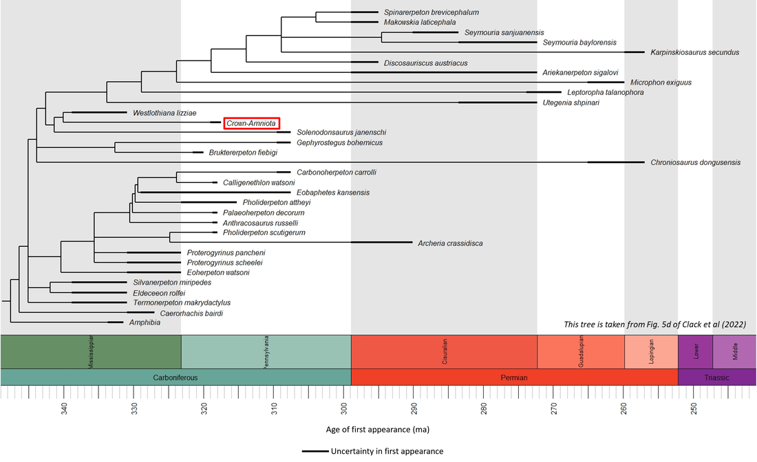
Figure 1. Phylogenetic time tree of the stem-Amniota
A number of fossils, all Early Carboniferous (Late Viséan) in age, can be considered as the oldest known representatives of the stem-Amniota:
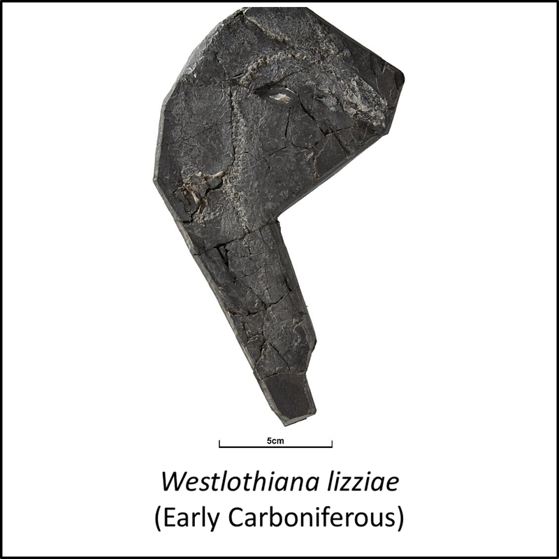
- Westlothiana lizziae
- Eldeceeon rolfei
- Silvanerpeton miripedes
- Termonerpeton makrydactylus


Names in red indicate that the fossil is younger than the oldest known crown-group fossil.
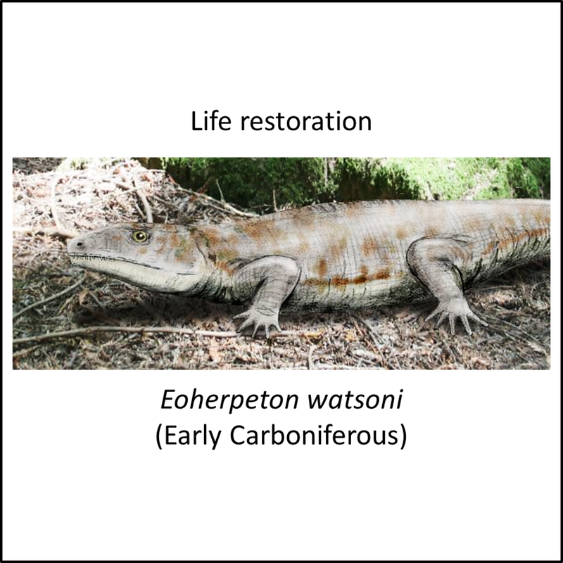
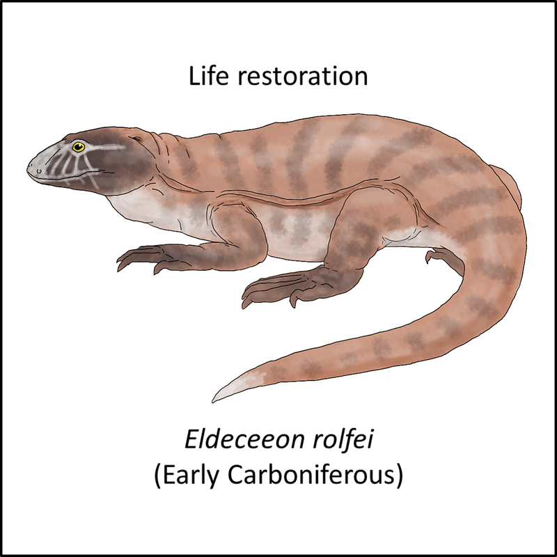
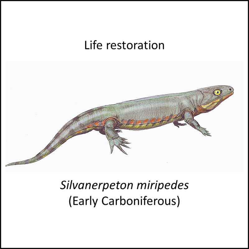
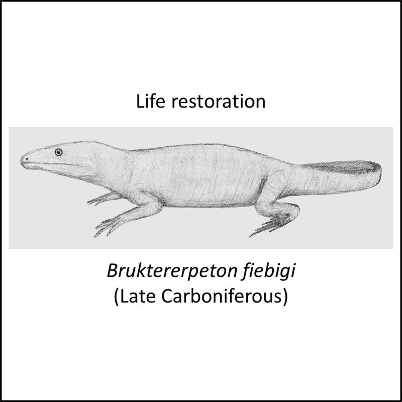
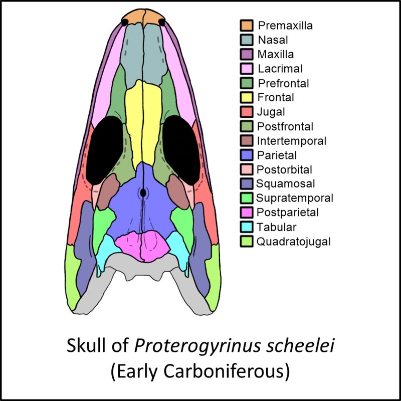
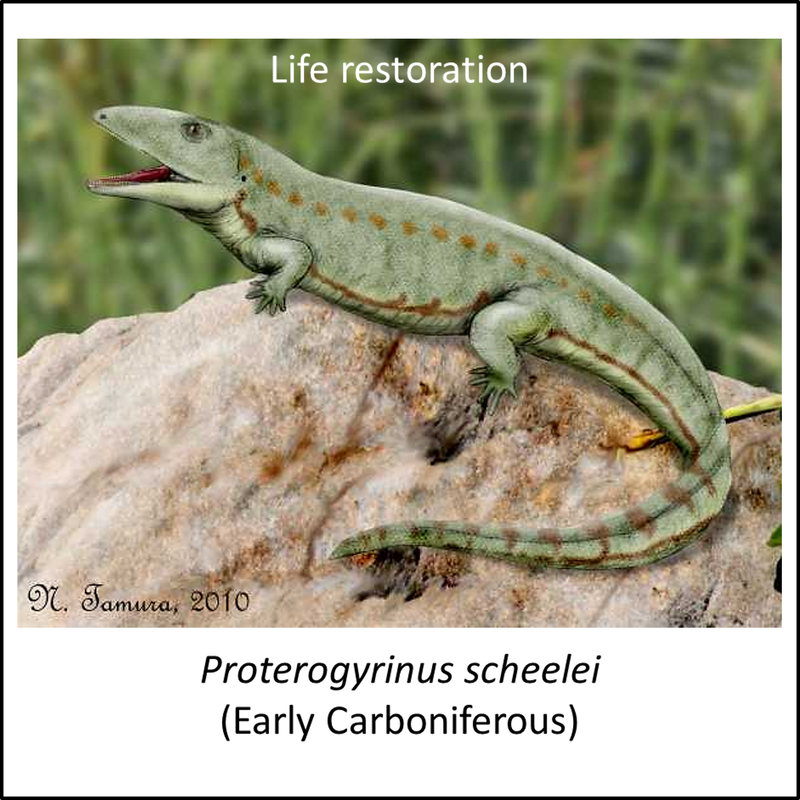
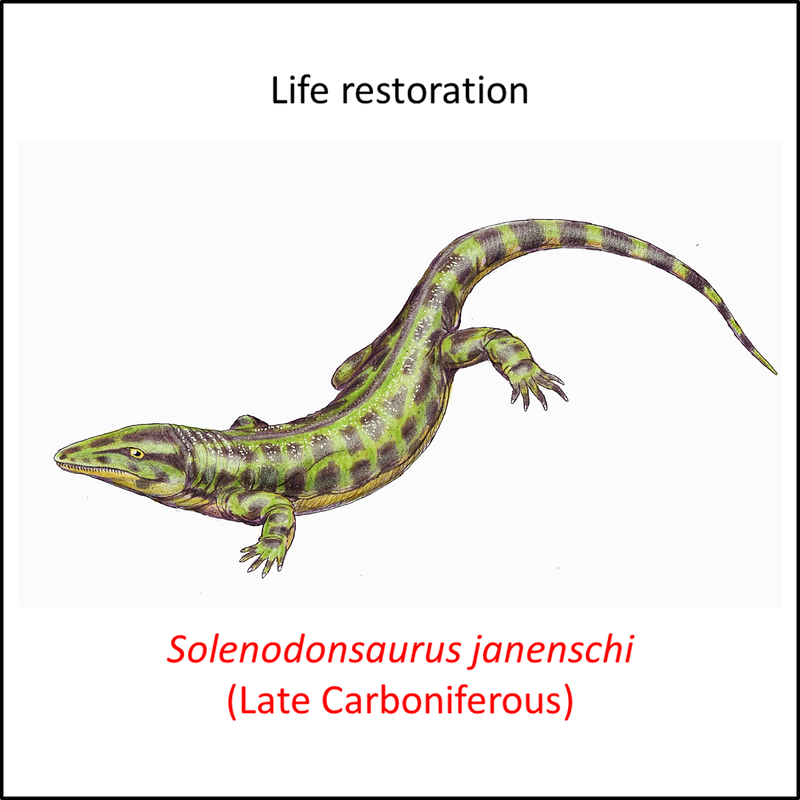
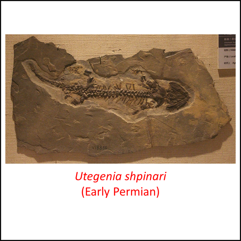
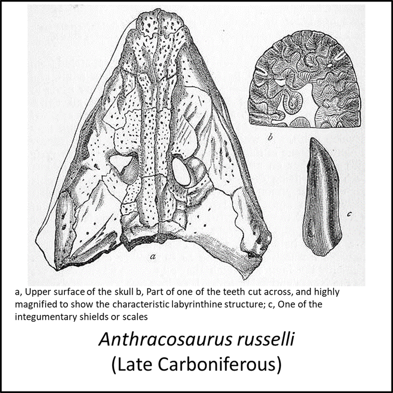
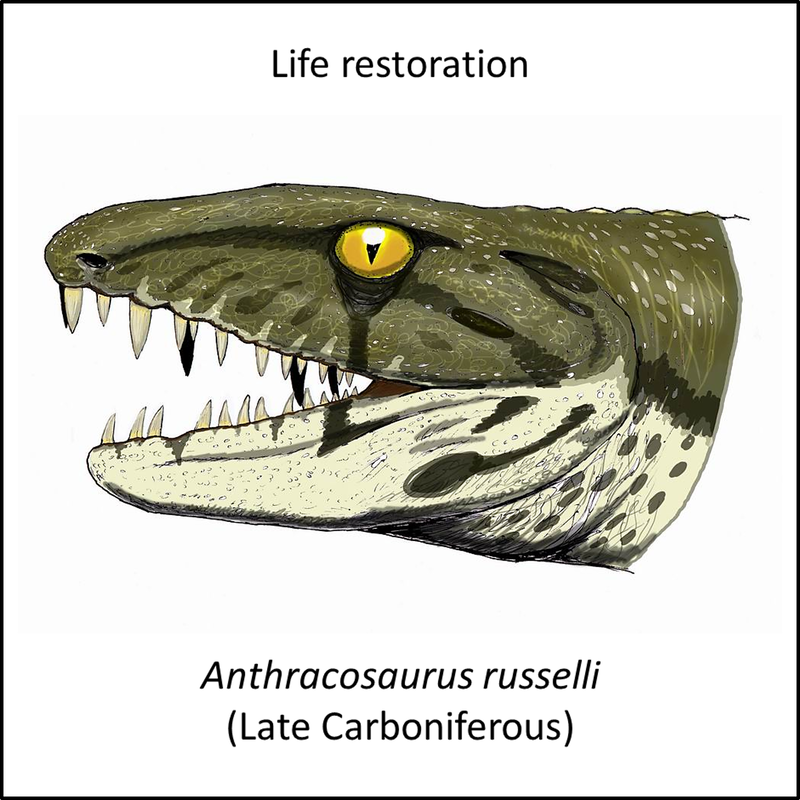
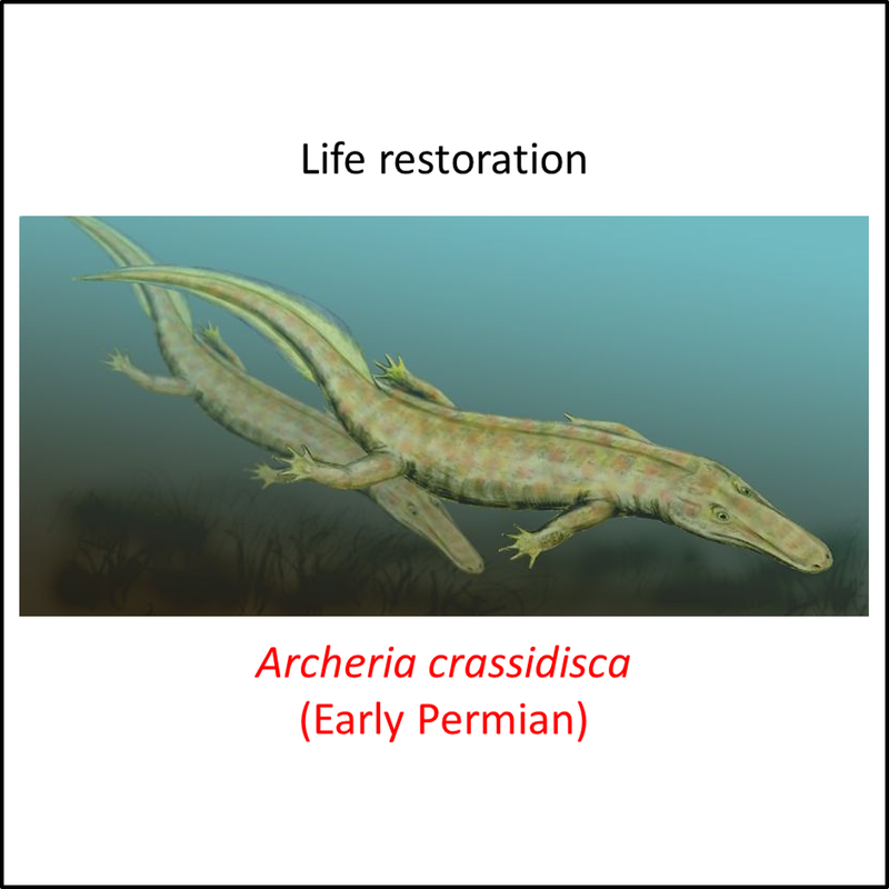
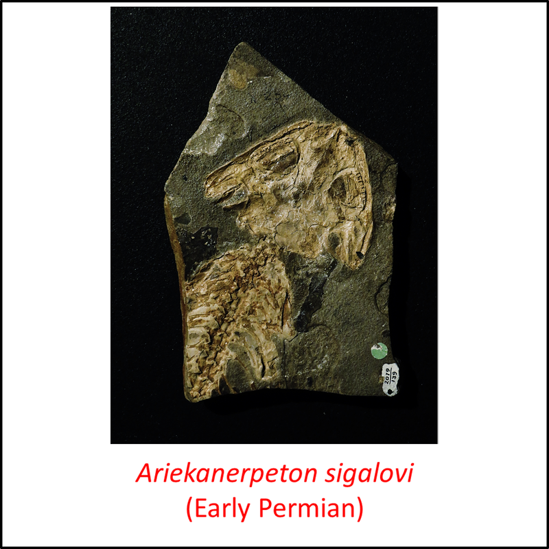
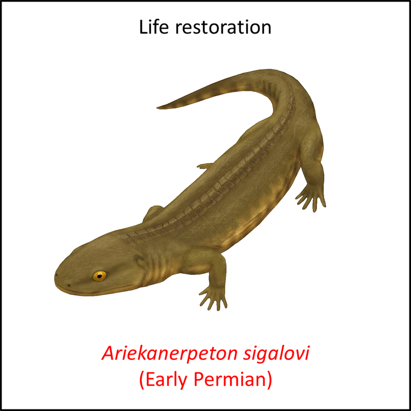
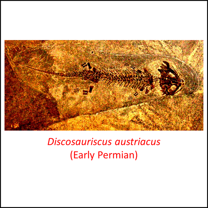
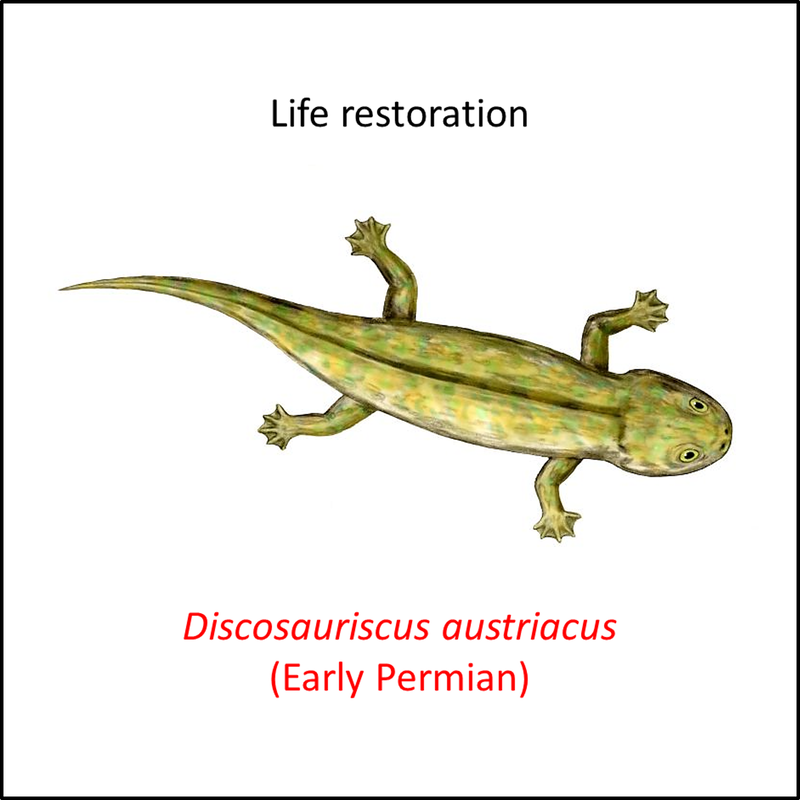
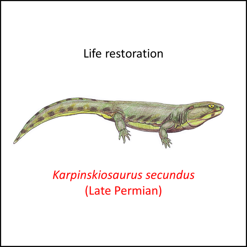
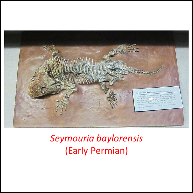
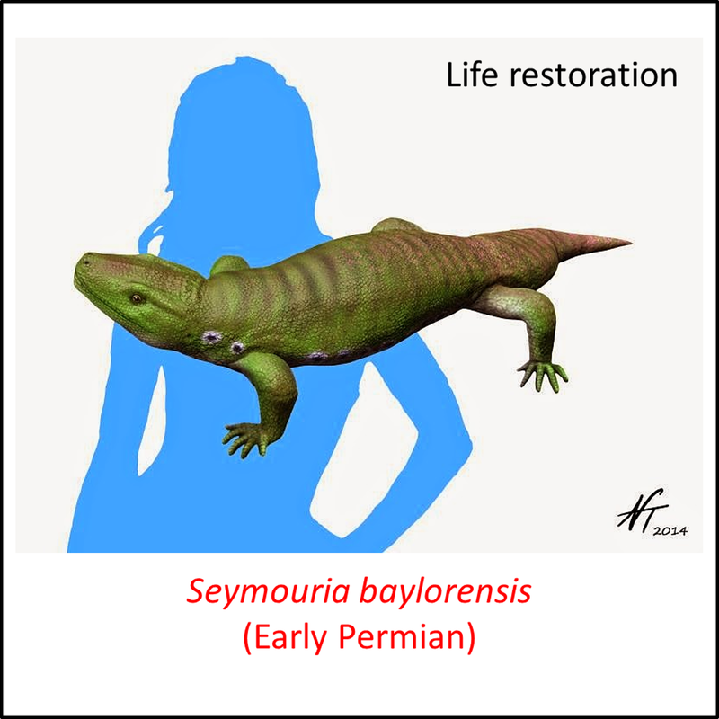
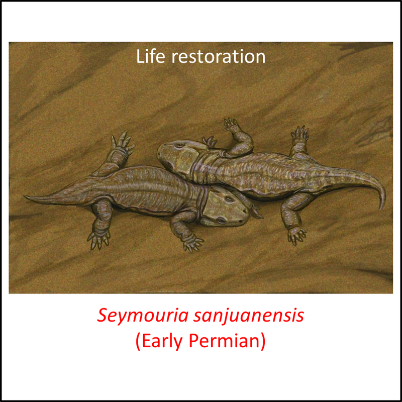
Figure 2. Images of stem-group amniotes
The above series of images illustrates some of the fossils that represent the ancestry of the amniote crown group. Note that most of the fossils (represented by names in red) post-date the appearance of the crown group; these represent branches of the stem line that continued to evolve after the crown-group had appeared.
The images are placed in order from most basal to most crownward in the stem group. There is no obvious change from the most basal to the most crownward, but there were in fact significant changes in jaw structure along the amniote stem line (Anderson et al, 2013).
Some idea of the nature of the transition from the stem group to the crown group of the amniotes can be obtained from a comparison of the above images with the examples of early crown-Amniota shown below:
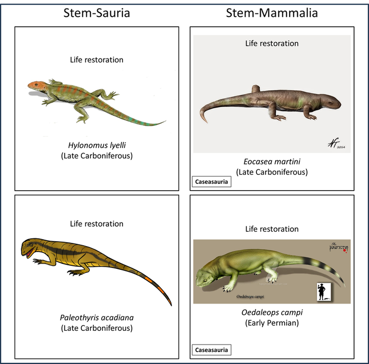
Figure 3. Examples of early crown-Amniota
The above time tree (Figure 1) indicates that the amniote stem group developed from Early Carboniferous to Late Permian time, but exclusion of the stem-group fossils that are younger than the oldest crown-group fossil indicates that the stem-to-crown transition was confined to the Carboniferous, as indicated below:

Figure 4. Rate of appearance of stem-group amniotes (predating the crown group and including only the genera shown in Figure 1)
Taking into account the fact that the oldest stem-group amphibian (Balanerpeton woodi) is a little older than the oldest known member of the stem-Amniota, the minimum duration of the amniote stem-to-crown transition is calculated to be about 12 million years.
References
Anderson, P. S. L., Friedman, M., & Ruta, M. (2013). Late to the Table: Diversification of Tetrapod Mandibular Biomechanics Lagged Behind the Evolution of Terrestriality. Integrative and Comparative Biology, Volume 53, Issue 2, 1 August 2013, p. 197–208.
Benton, M. J. (2015). Vertebrate Palaeontology - Fourth edition. John Wiley & Sons, 468 pages.
Clack, J. A., Smithson, T. R., & Ruta, M. (2022). A Mississippian (early Carboniferous) tetrapod showing early diversification of the hindlimbs. Nature communications biology, 5(1), 1-10.
Merck, J. (2015). The Amniotic Egg. Course notes for GEOL 431 Vertebrate Paleobiology, Spring Semester 2015, University of Maryland.
Schoch, R. R., & Sues, H. D. (2016). The diapsid origin of turtles. Zoology, 119(3), 159-161.
Image credits – Stem-Amniota
- Figure 2 (Caerorhachis bairdi): Богданов [email protected], Public domain, via Wikimedia Commons
- Figure 2 (Termonerpeton makrydactylus, fossil): Open Access article Clack, J. A., Smithson, T. R., & Ruta, M. (2022). A Mississippian (early Carboniferous) tetrapod showing early diversification of the hindlimbs. Communications biology, 5(1), 1-10.
- Figure 2 (Termonerpeton makrydactylus, life restoration): Nix Illustration, under under a Creative Commons Attribution-NonCommercial license (CC BY-NC 4.0)
- Figure 2 (Chroniosaurus dongusensis): Nobu Tamura, under Creative Commons Attribution-ShareAlike (CC BY-SA) license
- Figure 2 (Eoherpeton watsoni): Nobu Tamura, under Creative Commons Attribution-ShareAlike (CC BY-SA) license
- Figure 2 (Eldeceeon rolfei): Fanboyphilosopher (Neil Pezzoni), CC BY 3.0 <https://creativecommons.org/licenses/by/3.0>, via Wikimedia Commons
- Figure 2 (Silvanerpeton miripedes): Dmitry Bogdanov, CC BY-SA 3.0 <https://creativecommons.org/licenses/by-sa/3.0>, via Wikimedia Commons
- Figure 2 (Bruktererpeton fiebigi): Slate Weasel, CC0, via Wikimedia Commons
- Figure 2 (Gephyrostegus bohemicus): Dmitry Bogdanov, CC BY-SA 3.0 <https://creativecommons.org/licenses/by-sa/3.0>, via Wikimedia Commons
- Figure 2 (skull of Proterogyrinus scheelei): Fanboyphilosopher (Neil Pezzoni), CC BY 4.0 <https://creativecommons.org/licenses/by/4.0>, via Wikimedia Commons
- Figure 2 (Proterogyrinus scheelei, life restoration): Nobu Tamura, under Creative Commons Attribution-ShareAlike (CC BY-SA) license
- Figure 2 (Solenodonsaurus janenschi, fossil): Danto, M., Witzmann, F., and Müller, J., CC BY-SA 3.0 <https://creativecommons.org/licenses/by-sa/3.0>, via Wikimedia Commons
- Figure 2 (Solenodonsaurus janenschi, life restoration): Dmitry Bogdanov, CC BY 3.0 <https://creativecommons.org/licenses/by/3.0>, via Wikimedia Commons
- Figure 2 (Utegenia shpinari): Morosaurus millenii, CC BY-SA 4.0 <https://creativecommons.org/licenses/by-sa/4.0>, via Wikimedia Commons
- Figure 2 (Anthracosaurus russelli, fossil): H. ALLEYNE NICHOLSON, Public domain, via Wikimedia Commons
- Figure 2 (Anthracosaurus russelli, life restoration): Богданов (The original uploader was ДиБгд at Russian Wikipedia.), Public domain, via Wikimedia Commons
- Figure 2 (Archeria crassidisca): Nobu Tamura under a Creative Commons 3.0 Unported (CC BY-NC-ND 3.0) license
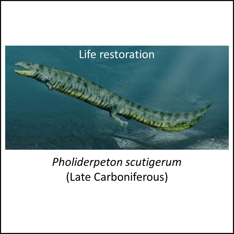
- Figure 2 (Pholiderpeton scutigerum): Nobu Tamura, under Creative Commons Attribution-ShareAlike (CC BY-SA) license
- Figure 2 (Westlothiana lizziae, fossil): British Geological Survey, under a Creative Commons Attribution-NonCommercial-ShareAlike 3.0 Unported License.
- Figure 2 (Westlothiana lizziae, life restoration): Nobu Tamura under a Creative Commons 3.0 Unported (CC BY-NC-ND 3.0) license
- Figure 2 (Leptoropha talanophora): Dmitry Bogdanov, CC BY-SA 3.0 <https://creativecommons.org/licenses/by-sa/3.0>, via Wikimedia Commons
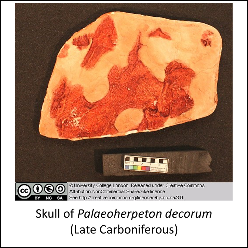
- Figure 2 (Palaeoherpeton decorum): University Collge London, under the Creative Commons Attribution Non-commercial Share Alike 3.0 License
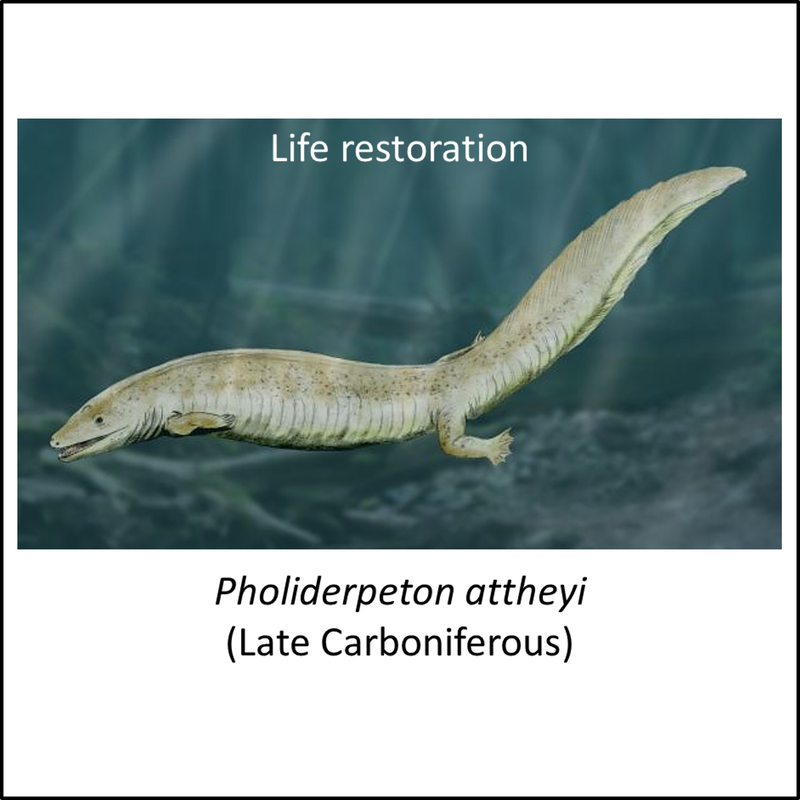
- Figure 2 (Pholiderpeton attheyi): Nobu Tamura, under Creative Commons Attribution-ShareAlike (CC BY-SA) license
- Figure 2 (Ariekanerpeton sigalovi, fossil): Ghedoghedo, CC BY-SA 4.0 <https://creativecommons.org/licenses/by-sa/4.0>, via Wikimedia Commons
- Figure 2 (Ariekanerpeton sigalovi, life restoration): Smokeybjb, CC BY-SA 3.0 <https://creativecommons.org/licenses/by-sa/3.0>, via Wikimedia Commons
- Figure 2 (Discosauriscus austriacus, fossil): GhedoghedoGhedoghedo, CC BY-SA 3.0 <https://creativecommons.org/licenses/by-sa/3.0>, via Wikimedia Commons
- Figure 2 (Discosauriscus austriacus, life restoration): Nobu Tamura, under Creative Commons Attribution-ShareAlike (CC BY-SA) license
- Figure 2 (Karpinskiosaurus secundus): Dmitry Bogdanov, CC BY-SA 3.0 <https://creativecommons.org/licenses/by-sa/3.0>, via Wikimedia Commons
- Figure 2 (Seymouria baylorensis, fossil): Greg Goebel from Loveland CO, USA, CC BY-SA 2.0 <https://creativecommons.org/licenses/by-sa/2.0>, via Wikimedia Commons
- Figure 2 (Seymouria baylorensis, life restoration): Nobu Tamura, under Creative Commons Attribution-ShareAlike (CC BY-SA) license
- Figure 2 (Seymouria sanjuanensis, fossils): James St. John, CC BY 2.0 <https://creativecommons.org/licenses/by/2.0>, via Wikimedia Commons
- Figure 2 (Seymouria sanjuanensis, life restoration): Dmitry Bogdanov, CC BY 3.0 <https://creativecommons.org/licenses/by/3.0>, via Wikimedia Commons
- Figure 2 (Makowskia laticephala): J. Klembara, CC BY-SA 4.0 <https://creativecommons.org/licenses/by-sa/4.0>, via Wikimedia Commons
- Figure 3 (Hylonomus lyelli): Nobu Tamura under a Creative Commons 3.0 Unported (CC BY-NC-ND 3.0) license
- Figure 3 (Paleothyris acadiana): Conty, CC BY-SA 4.0 <https://creativecommons.org/licenses/by-sa/4.0>, via Wikimedia Commons
- Figure 3 (Eocasea martini): Nobu Tamura, licensed under Creative Commons Attribution- ShareAlike (CC BY-SA) license
- Figure 3 (Oedaleops campi): Theropsida, Creative Commons Attribution-NonCommercial-NoDerivs 3.0 Unported (CC BY-NC-ND 3.0)