The hyraxes or dassie (Order Hyracoidea, Infraclass Eutheria) are small hoofed mammals native to Africa and extreme southwestern Asia that are currently known in six extant species (Encyclopaedia Britannica). These species all belong to the family Procaviidae.
The most extensive phylogenetic analysis available for the hyracoid stem group is that presented by Puttick and Thomas (2015). This analysis is the basis for the following phylogenetic time tree:
The most extensive phylogenetic analysis available for the hyracoid stem group is that presented by Puttick and Thomas (2015). This analysis is the basis for the following phylogenetic time tree:
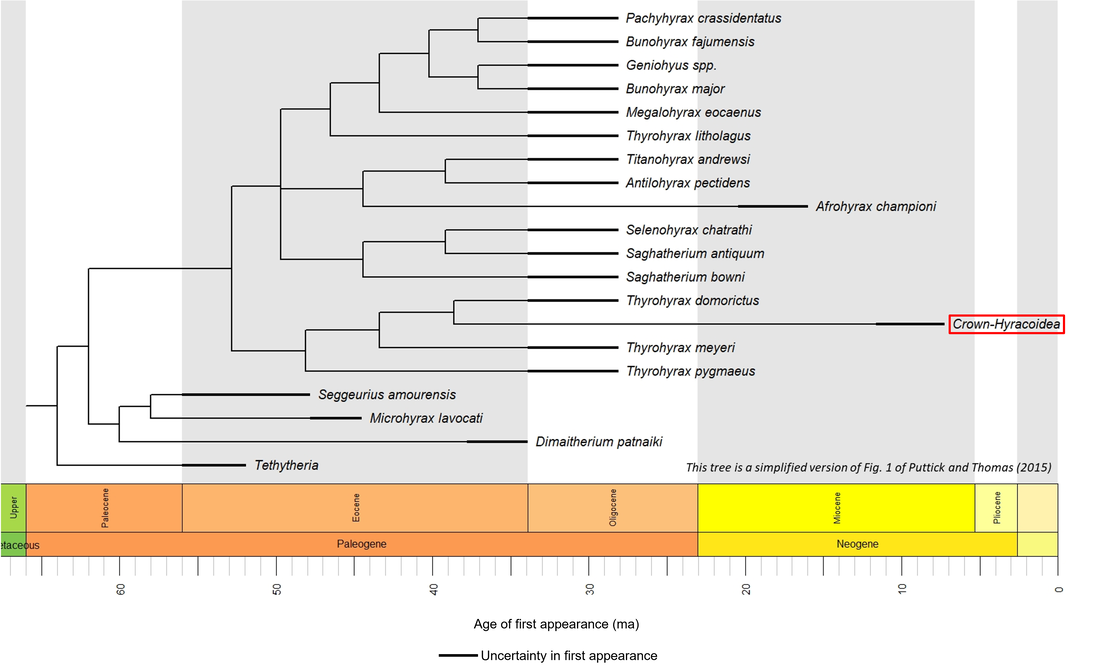
Figure 1. Time tree of the stem-Hyracoidea
The oldest known member of the stem-Hyracoidea is Seggeurius amourensis, described from the Early Eocene (Ypresian) El Kohol Formation in the Southern Atlas of Algeria (Mahboubi, 1993; Heritage et al, 2021). Unfortunately, no public-domain image is available either of this species or of most of the other stem-group fossils represented in the above time tree. The few available ones are shown below (for a larger view, click on image):
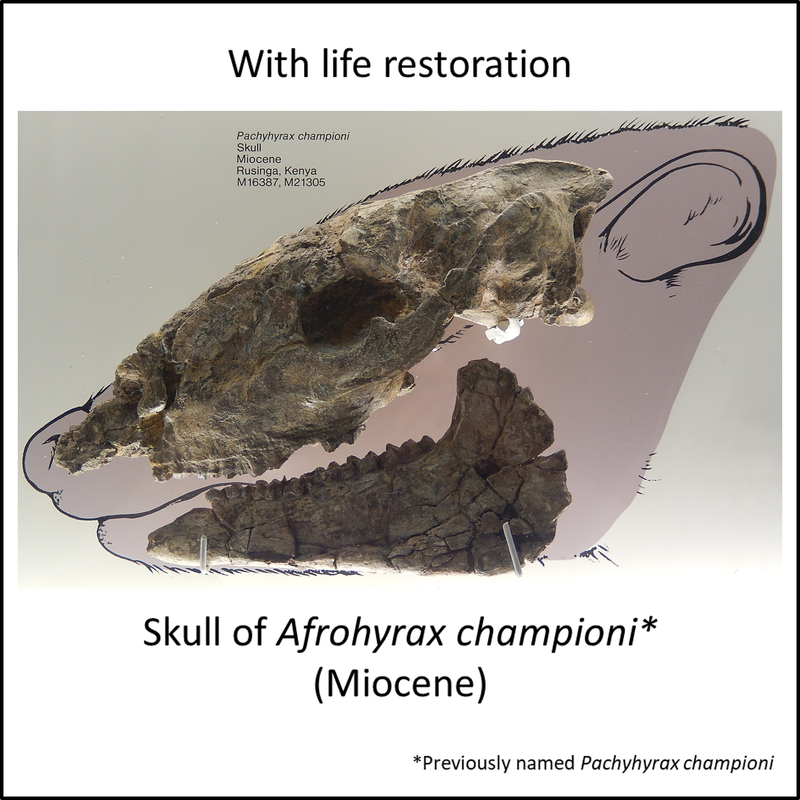
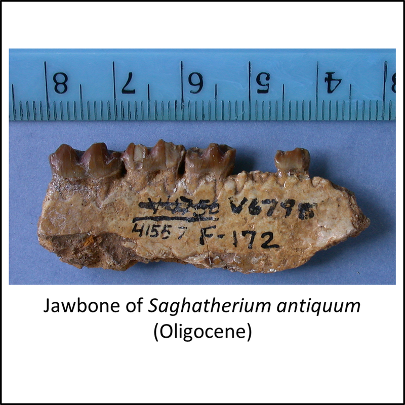

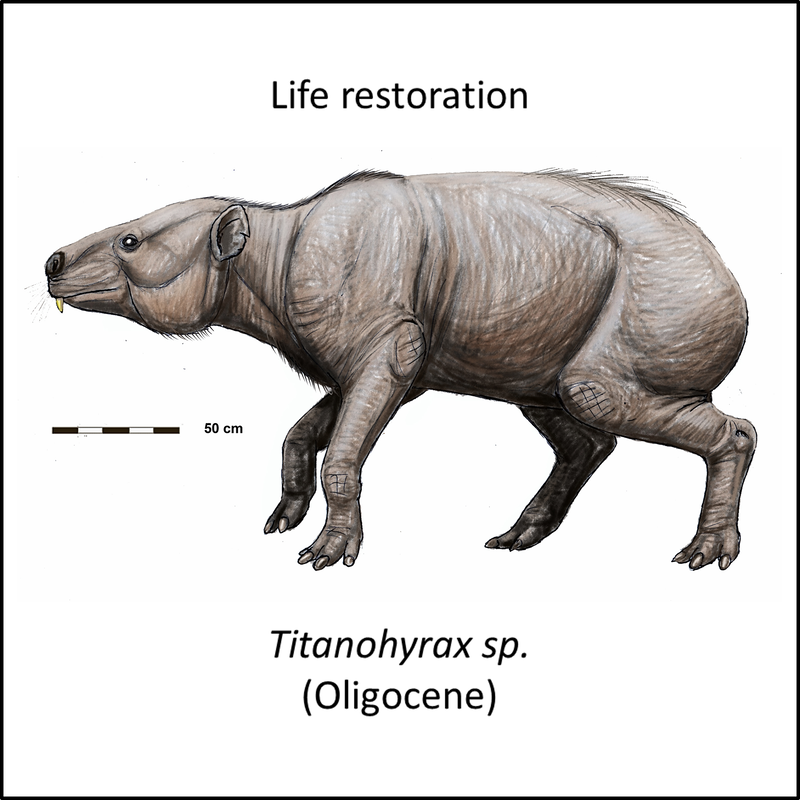
Figure 2. Images of stem-Hyracoidea
The oldest known member of the hyracoid crown group is Heterohyrax auricampensis, described from a Late Miocene (Tortonian) fissure fill breccia at Berk Aukas (Site I) in Namibia (Heritage et al, 2021). Again, no image is available in the public domain.
The available fossil data indicate that the hyracoid stem group developed from Early Eocene to Late Miocene time, representing a stem-to-crown transition of at least 36 million years (see Figure 1). As shown below, the rate of evolutionary change was highest during the Oligocene:
The available fossil data indicate that the hyracoid stem group developed from Early Eocene to Late Miocene time, representing a stem-to-crown transition of at least 36 million years (see Figure 1). As shown below, the rate of evolutionary change was highest during the Oligocene:
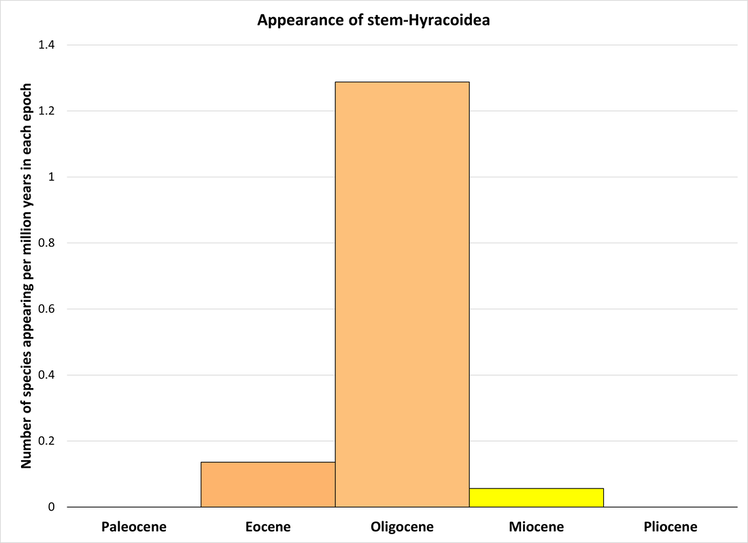
Figure 3. Rate of appearance of stem-group hyraxes (predating the crown group and including only the species shown in Figure 1)
References
Barrow, E. C., Seiffert, E. R., & Simons, E. L. (2012). Cranial morphology of Thyrohyrax domorictus (Mammalia, Hyracoidea) from the early Oligocene of Egypt. Journal of Vertebrate Paleontology, 32(1), 166-179.
Heritage, S., Seiffert, E. R., & Borths, M. R. (2021). Recommended fossil calibrators for time-scaled molecular phylogenies of Afrotheria. Afrotherian Conservation 17 https://www. afrotheria. net/newsletter. php.
Mahboubi, M. (1993). Reassessment of lower Eocene Seggeurius amourensis: aspects of primitive dental morphology in the mammalian order Hyracoidea. Journal of Paleontology, 67(5), 889-893.
Puttick, M. N., & Thomas, G. H. (2015). Fossils and living taxa agree on patterns of body mass evolution: a case study with Afrotheria. Proceedings of the Royal Society B: Biological Sciences, 282(1821), 20152023.
Image credits – stem-Hyracoidea
- Header (Rock hyrax, Procavia capensis, photographed in Israel): Greg Schechter from San Francisco, USA, CC BY 2.0 <https://creativecommons.org/licenses/by/2.0>, via Wikimedia Commons
- Figure 2 (Afrohyrax championi): Chiswick Chap, earlier version Ghedoghedo, CC BY-SA 3.0 <https://creativecommons.org/licenses/by-sa/3.0>, via Wikimedia Commons
- Figure 2 (Saghatherium antiquum): Regents of the University of California, under Creative Commons Attribution 3.0 (CC BY 3.0) license
- Figure 2 (Titanohyrax andrewsi): Gertrude Mary Woodward (1861-1939), CC BY-SA 4.0 <https://creativecommons.org/licenses/by-sa/4.0>, via Wikimedia Commons
- Figure 2 (Titanohyrax sp.): DiBgd, CC BY-SA 4.0 <https://creativecommons.org/licenses/by-sa/4.0>, via Wikimedia Commons